INTRODUCTION
Antibiotic-associated diarrhea (AAD) is a common gastrointestinal adverse reaction associated with the clinical use of broadspectrum antibiotics, and it is particularly prevalent among patients receiving antibiotic therapy [Zhang et al., 2022]. AAD not merely affects patients’ quality of life and treatment adherence, but may lead to complications such as dehydration, electrolyte imbalances, and Clostridioides difficile infection [Abad & Safdar, 2021]. Existing research indicates that the core pathogenesis of AAD lies in the “indiscriminate attack” of antibiotics on the gut microbiota, leading to an imbalance in the intestinal microecology and metabolic dysfunction [Ramirez et al., 2020]. Specifically, the amount of short-chain fatty acids (SCFAs), such as acetic and propionic acids, generated by gut microbial fermentation of dietary fiber, decreases significantly. Therefore, identifying intervention strategies that can effectively modulate the gut microbiota and restore SCFA levels is crucial for the prevention and treatment of AAD.
The gut microbiota, regarded as the body’s “forgotten organ”, serves a key function in preserving the health of the host [Wei et al., 2024]. A close metabolic relationship exists between the gut microbiota and SCFAs. On the one hand, the gut microbiota secretes various carbohydrate-active enzymes to break down dietary fiber and polysaccharides, which are difficult for the host to digest, into monosaccharides or oligosaccharides, and then converted into SCFAs through metabolic pathways such as glycolysis and the pentose phosphate pathway. On the other hand, SCFAs, as key metabolic products of the microbiota, reciprocally regulate the composition and function of the gut microbiota, forming an interactive regulatory network among the microbiota, metabolites, and host [Hays et al., 2024]. SCFAs exert a wide range of physiological effects: acetic acid, the most abundant shortchain fatty acid, regulates fat metabolism and appetite; propionic acid contributes to immune regulation and alleviates intestinal inflammation; butyric acid serves as the primary energy source for intestinal epithelial cells, promotes the expression of tight junction proteins, enhances intestinal barrier function, and maintains intestinal immune homeostasis. Accordingly, maintaining the dynamic balance between the gut microbiota structure and SCFAs metabolism is vital for the prevention and treatment of intestinal diseases.
In recent years, polysaccharides derived from medicinal and edible plants have demonstrated significant potential in modulating the gut microbiota due to their favorable safety profiles and notable prebiotic activity [Huang et al., 2023; Liu et al., 2024; Xu et al., 2023]. These polysaccharides are typically resistant to degradation by human digestive enzymes but can be selectively utilized by the gut microbiota. Polygonatum sibiricum Red., a traditional and valuable edible-medicinal herb in China, has been extensively studied; its rhizomes have been shown to possess immunomodulatory, anti-inflammatory, and gut microbiota-regulating properties, with its polysaccharides considered the primary source of these bioactivities [Kuang et al., 2024; Lai et al., 2024]. However, during the cultivation and processing of Polygonatum, its aerial parts (leaves) are routinely discarded as waste, resulting in a significant waste of resources. Phytochemical studies have shown that Polygonatum leaves are also rich in polysaccharides, and their polysaccharides composition differs from that of the rhizomes, suggesting they may possess unique biological activities [Li et al., 2022]. One should note that Polygonatum leaves have a history of culinary use in certain folk traditions across China; nevertheless, modern pharmacological research on them remains insufficient, particularly regarding their potential for regulating the gut microbiota and preventing or treating intestinal diseases. Given that the gut microbiota serves as a key bridge between dietary components and host health, studies on its response characteristics and metabolic transformation patterns regarding the P. sibiricum leaf polysaccharides (PsPs) in intestinal diseases are lacking.
Our previous research has shown that PsPs can increase the proportion of Firmicutes and reduce the abundance of Bacteroidetes in the gut microbiota of healthy mice, while significantly increasing SCFAs production [Luo et al., 2022]. These results indicate that PsPs are promising candidates for prebiotic formulation development. Building on this, this study further investigates the alleviating effects of PsPs on AAD. We first extracted PsPs via water extraction and ethanol precipitation, and then evaluated their stability in the gastrointestinal environment, as well as their effects on microbial community structure and SCFAs production, through in vitro simulated digestion and human fecal fermentation experiments. Thereafter, the PsPs were evaluated for their effects on diarrhea symptoms, colonic histopathology and intestinal microbiota profile in C57BL/6J mice with AAD induced by lincomycin hydrochloride. The purpose of this study was to elucidate the beneficial effects of PsPs on AAD, with the goal of providing scientific evidence to support the high-value utilization of Polygonatum leaves (a non-traditional medicinal part), and offering new insights and experimental support for the development of AAD adjunctive intervention strategies based on polysaccharides derived from food-medicine dual-use sources.
MATERIALS AND METHODS
Materials and chemicals
Healthy mature P. sibiricum leaves harvested in Chongqing, China. Standard SCFAs were purchased from Sigma-Aldrich (Shanghai, China). Dextran standards were purchased from Aladdin Biochemical Technology Co., Ltd. (Shanghai, China). The reducing sugar assay kit was purchased from Solarbio Technology Co., Ltd. (Beijing, China). The total antioxidant capacity assay kit was purchased from Beyotime Biotechnology Co., Ltd. (Shanghai, China). The inflammatory cytokine assay kits used to determine monocyte chemoattractant protein-1 (MCP-1), lipopolysaccharide (LPS), interleukin-6 (IL-6), and tumor necrosis falpha (TNF-α) were purchased from ZCIBIO Technology Co. Ltd. (Shanghai, China). Anti-zonula occludens-1 (ZO-1) antibody was purchased from Wuhan Servicebio Co., Ltd. (Wuhan, China). The anti-zoocadin-1 and anti-zoocadin-2 antibodies were supplied by Proteintech Group Co., Ltd. (Chicago, IL, USA). The AnaeroPack anaerobic culture bags were purchased from Mitsubishi Chemical Analytech Co. Ltd (Tokyo, Japan).
Separation of leaf polysaccharides and their characterization
PsPs were obtained by extracting P. sibiricum leaves with water and precipitating with ethanol according to the method described in our previous study [Luo et al., 2022]. Crushed leaves (2.5 kg) were soaked in water at twice their volume and heated in a 90°C water bath for 3 h. The extraction was repeated three times. Then anhydrous ethanol at 1.5 volumes of the extraction liquid was added. After 24 h room-temperature standing, the solution was filtered to obtain crude polysaccharides. Afterward, the crude polysaccharides were dissolved in distilled water and decolorized by the macroporous resin. The crude polysaccharides solution was further purified using diethylaminoethyl (DEAE) cellulose, and then dialyzed in a dialysis bag (0.5–1.0 kDa) for 72 h. After dialysis, the purified polysaccharides were concentrated and freeze-dried to yield the dry powder. The monosaccharide composition of PsPs was: rhamnose (15.4%), arabinose (24%), glucose (8.8%), xylose (40.7%), mannose (6.6%), and galacturonic acid (4.5%) [Luo et al., 2022]. The average molecular weight of PsPs was determined by high-performance gel-permeation chromatography (HPGPC) using an LC-2010 HPLC system (Shimadzu Corporation, Kyoto, Japan) with refractive index (RI) detector, equipped with a TSK-Gel GMPWXL column (7.8×300 mm, particle diameter 13 μm; Thermo Fisher Scientific, Waltham, MA, USA). The mobile phase consisted of a 0.02 M KH2PO4 solution. PsPs solutions (5 mg/mL) were filtered through a 0.22 μm filter membrane. Next, 20 μL of the filtered sample were injected into the HPLC column. A standard curve was prepared using dextran standards (1, 2, 30, 40, 70, 100, and 200 kDa). The average molecular weight of PsPs was calculated based on the calibration curve plotted using these dextran standards. The surface features and microstructure of the PsPs were characterized in detail using a SIGMA 300 scanning electron microscope (SEM; ZEISS, Oberkochen, Germany). Before analysis, the samples were given a gold coating under reduced pressure to improve conductivity and imaging quality. SEM photographs were taken at ×100 magnification.
In vitro models assessing leaf polysaccharides stability and microbial utilization
The in vitro models assessing PsPs stability and microbial utilization were conducted using modified versions of previously reported methods [Ma et al., 2025; Zhao et al., 2023]. PsPs (10 mg/mL) was dissolved in simulated saliva (primarily including NaCl, NaHCO3, K2CO3; pH 6.8). Then equal volumes of simulated gastric (primarily including HCl, NaCl, pepsin; pH 1.5) and intestinal fluids (primarily including KH2PO4, K2HPO4, NaOH; pH 7.8) were added, and the mixture was incubated with shaking at 37°C to simulate the digestive process. At predetermined time points (0, 2 and 5 min) of salivary digestion, as well as 0.5, 1, 2 and 4 h of oral-gastric and oral-gastrointestinal digestion respectively, samples were collected for testing. The reaction mixture was heated in boiling water for 6 min. Heat treatment inactivated the digestive enzymes, thereby terminating the digestion. Subsequently, the total antioxidant capacity and reducing sugar content of the digested samples were determined employing commercial kits (Beyotime Biotechnology Co., Ltd. and Solarbio Technology Co., Ltd., respectively) and the microplate reader, and the total sugar content was measured using the phenolsulfuric acid method with spectrophotometry [Dubois et al., 1956]. The results of total antioxidant capacity were expressed as the amount of the antioxidant Trolox equivalent contained in each liter of solution. The results of reducing sugar content were expressed as the amount of mass of reducing sugars per milliliter of digesta solution. The results of total sugar content were expressed as the percentage by PsPs weight (%, w/w).
Fresh fecal samples were collected from three healthy male volunteers (aged 20–25 years) who had not taken antibiotics in the past three months, and the samples were mixed in equal volumes. Phosphate-buffered saline (PBS) was used to prepare a 10% (w/v) fecal suspension, which was then centrifuged (4°C, 908×g, 5 min). The resulting supernatant was blended with Luria-Bertani medium (without sugar source) containing PsPs (10 mg/mL) at a 1:9 (v/v) ratio to form the PsPs group. The blank control group (CN) was prepared in the same proportion without the addition of PsPs. The preparations were placed in AnaeroPack anaerobic culture bags. Anaerobic fermentation was conducted at 37°C for 24 h under conditions where oxygen concentration was below 0.1% and carbon dioxide concentration was maintained at approximately 10%. Finally, fermentation broths at 0, 6, 12, and 24 h were collected and centrifuged (4°C, 12,880×g, 3 min) to separate bacterial pellets from the fermentation supernatant for further microbial community and SCFAs analysis.
Animal experiments
Five-week-old male C57BL/6J mice were housed for 7 days in a standard facility (12 h light-dark cycle, 23±0.5°C, relative humidity 50±5%) to allow adaptation before the start of gavage administration. Thirty-two mice were randomly divided into four groups (n=8): AAD group, blank control group (CN), PsPs group, and natural recovery group (NR). The experiment involved continuous gavage for 10 days [Lai et al., 2025]. CN group received twice-daily gavage of normal saline for 10 days. Remaining three groups were gavaged with 3 g/kg lincomycin hydrochloride daily twice for three days to induce AAD. Afterward, the PsPs group was gavaged with 300 mg/kg PsPs daily twice a day, and the NR group was gavaged daily with normal saline twice a day, for 7 days. Finally, the cecum, blood, and feces of four groups mice were collected for subsequent experimental analysis. All experiments were approved by the Ethics Committee of Southwest Medical University (SWMU, Luzhou, China), approval number swmu20250266.
16S ribosomal RNA sequencing and bioinformatics analysis
DNA extraction was performed on specimens derived from microbial fermentation and on mouse fecal matter with the QIAamp fast DNA stool mini kit (Qiagen, Hilden, NRW, DEU). Genomic DNA served as the template for PCR amplification of the 16S rRNA V3-V4 region, using the primers 338-F (5′-ACTCCTACGGGAGGCAGCAG-3′) and 806-R (5′-GGACTACHVGGGTWTCTAAT-3′) [Xu et al., 2016]. And then, the PCR products were subjected to high-throughput sequencing at MajorBio Bio-Pharm Technology Co., Ltd. (Shanghai, China). We utilized the MajorBio cloud platform (https://cloud.majorbio.com) to analyze the sequencing data. Quality control was performed on the sequencing reads, and they were assigned to amplicon sequence variant (ASV) clusters using a 100% similarity threshold. The identified ASVs were mapped to the Silva (SSU123) 16S rRNA database using the RDP classification algorithm (http://rdp.cme.msu.edu) to analyze their taxonomic characteristics. Alpha diversity was calculated using the MOTHUR software (version v.1.30.2; Patrick Schloss, Ann Arbor, MI, USA). Higher values of abundance-based coverage estimator (ACE) indicate greater species diversity, higher Shannon indices indicate higher species evenness, and higher Simpson indices indicate higher species concentration. A hierarchical clustering tree was constructed using the Bray-Curtis index via the Qiime2 software (Rob Knight, San Diego, CA, USA), followed by principal co-ordinates analysis (PCoA). The raw sequencing data and alignment information for the experimental samples are stored in the NCBI Sequence Read Archive (accession number: PRJNA1419527).
Short-chain fatty acid determination
The SCFA content in the supernatant of the fermentation broth was quantified using liquid chromatography–mass spectrometry (LC–MS/MS). Sample analysis was performed using an Exion-LC AD liquid chromatography system coupled with a QTRAP® 6500+ mass spectrometer (AB Sciex, Framingham, MA, USA). The procedure involved injecting 2 μL of the sample onto a BEH C18 (150×2.1 mm, 1.7 μm; Waters, Milford, MA, USA) column for chromatographic separation, followed by mass spectrometric detection. The mobile phase was composed of two solvents: A (water with 0.1% formic acid, v/v) and B (acetonitrile with 0.1% formic acid, v/v). Separation was achieved within 16 min at 40°C and a flow rate of 0.35 mL/min. The SCFAs were identified based on standards and literature references, and their content was quantified using a standard curve. The results were expressed as the mass of SCFA per milliliter of fermentation broth.
Tissue staining and immunofluorescence analysis
After formaldehyde fixation and paraffin embedding, cecal tissue sections were deparaffinized in xylene and rehydrated in ethanol. To examine histopathological changes, cecal sections were stained with hematoxylin and eosin (HE).
Cecal tissue sections were first fixed with formaldehyde and then washed with 100% ethanol. After incubation in a repair buffer and blocking with bovine serum albumin, the sections were incubated overnight at 4°C with ZO-1 antibody and antioccludin-1 antibody. After incubation with Cy3-labeled ZO-1 antibody and FITC-labeled occludin-1 antibody, the sections were analyzed using an immunofluorescence microscope (Nikon, Tokyo, Japan).
Inflammatory factor measurement
The enzyme-linked immunosorbent assay (ELISA) kit (ZCIBIO Technology Co. Ltd.) was performed to determine the inflammatory cytokines profiles including MCP-1, LPS, IL-6, and TNF-α in mouse serum. After blood collection from the mice’s eyes, the samples were kept stationary and then centrifuged to separate the supernatant serum, and the inflammatory factor concentrations were measured using the assay kit on the microplate reader (450 nm). The results of inflammatory factor concentrations were expressed as the amount of inflammatory cytokine per milliliter of serum.
Statistical analysis
Results from in vitro experiments were presented as the mean and standard deviation (SD) from three independent experiments. Results from animal experiments were expressed as the mean and SD from independent experiments of 8 mice per group. GraphPad Prism (v. 10.1.2; GraphPad Software, San Diego, CA, USA) and SPSS (v. 26; IBM, Armonk, New York, USA) were employed for statistical evaluation. Multigroup comparisons of body weight, colon length, and cytokine levels in animal experiments were performed using one-way analysis of variance (ANOVA). The t-test was used to analyze the results from the in vitro fermentation or to compare specific abundances between two groups. The α-diversity indices ACE, Shannon index and Simpson index were used the Kruskal–Wallis rank-sum test.
RESULTS AND DISCUSSION
Characterization of leaf polysaccharides
The average molecular weight of PsPs was 1.403±0.056×10⁵ Da. SEM disclosed the microstructure of PsPs (Figure S1 in Supplementary Materials). PsPs exhibits a flat, smooth, and porous lamellar morphology, which may allow it to be more effectively decomposed and utilized by the gut microbiota.
In vitro simulated digestion of leaf polysaccharides
As displayed in Table 1, the total sugar content of PsPs did not undergo significant changes (p≥0.05) during the simulated digestion period, indicating that PsPs remained largely stable and was not extensively degraded under the simulated digestive conditions. The reducing sugar content of PsPs increased sharply upon initial contact with gastric digestive fluid and then gradually decreased (Table 1). The increase was likely due to the highly acidic environment of gastric fluid breaking some of the glycosidic bonds in PsPs, thus exposing more reducing ends on the sugar chains. This result is consistent with findings in the literature indicating that gastric stimulation affects reducing sugar content [Wu et al., 2021]. In Table 1, the total antioxidant capacity of PsPs gradually decreases during digestion until it tends to stabilize.
Table 1
Total sugar content, reducing sugar content, and total antioxidant capacity during in vitro digestion of Polygonatum sibiricum Red. leaf polysaccharides (PsPs).
In vitro simulated fermentation of leaf polysaccharides
The changes in total antioxidant capacity during the PsPs fermentation period are shown in Table 2. No significant (p≥0.05) changes were observed during the first 6 h, followed by a continuous, significant (p<0.05) decrease. Results in Table 2 show that the reducing sugar content in PsPs did not change significantly (p≥0.05) during the first 6 h of fermentation but began to decrease markedly at 12 h. This could be attributed to the ongoing fermentation, during which the sugar chains were further degraded for microbial consumption, leading to a decrease of reducing sugar content in the later fermentation phases. The above results indicate that PsPs may be utilized by microbial communities during the middle to late stages of in vitro fermentation.
Table 2
Total antioxidant capacity and reducing sugar content of broths during in vitro fermentation of Polygonatum sibiricum Red. leaf polysaccharides (PsPs).
| Parameter | 0 h | 6 h | 12 h | 24 h |
|---|---|---|---|---|
| Total antioxidant capacity (mmol TE/L) | 0.78±0.04a | 0.76±0.02a | 0.67±0.02b | 0.60±0.01c |
| Reducing sugar content (mg/mL) | 0.70±0.04a | 0.88±0.10a | 0.43±0.07b | 0.30±0.02b |
The pH of the fermentation broth was recorded at specific time points (Figure 1A). In the unfermented state, no significant (p≥0.05) difference was observed between the PsPs and the CN group. During fermentation, the pH of the PsPs decreased at faster rates than the CN group.
Figure 1
In vitro fermentation of PsPs. (A) Changes in pH during fermentation. (B) Profile of short-chain fatty acids (SCFAs) after fermentation. (C) and (D) The proportions in the abundance of bacteria at the phylum and genus level, respectively. CN, blank control group; PsPs, Polygonatum sibiricum Red. leaf polysaccharides group. *, **, ***, and ****indicate statistical significance at p<0.05, p<0.01, p<0.001, and p<0.0001, respectively.
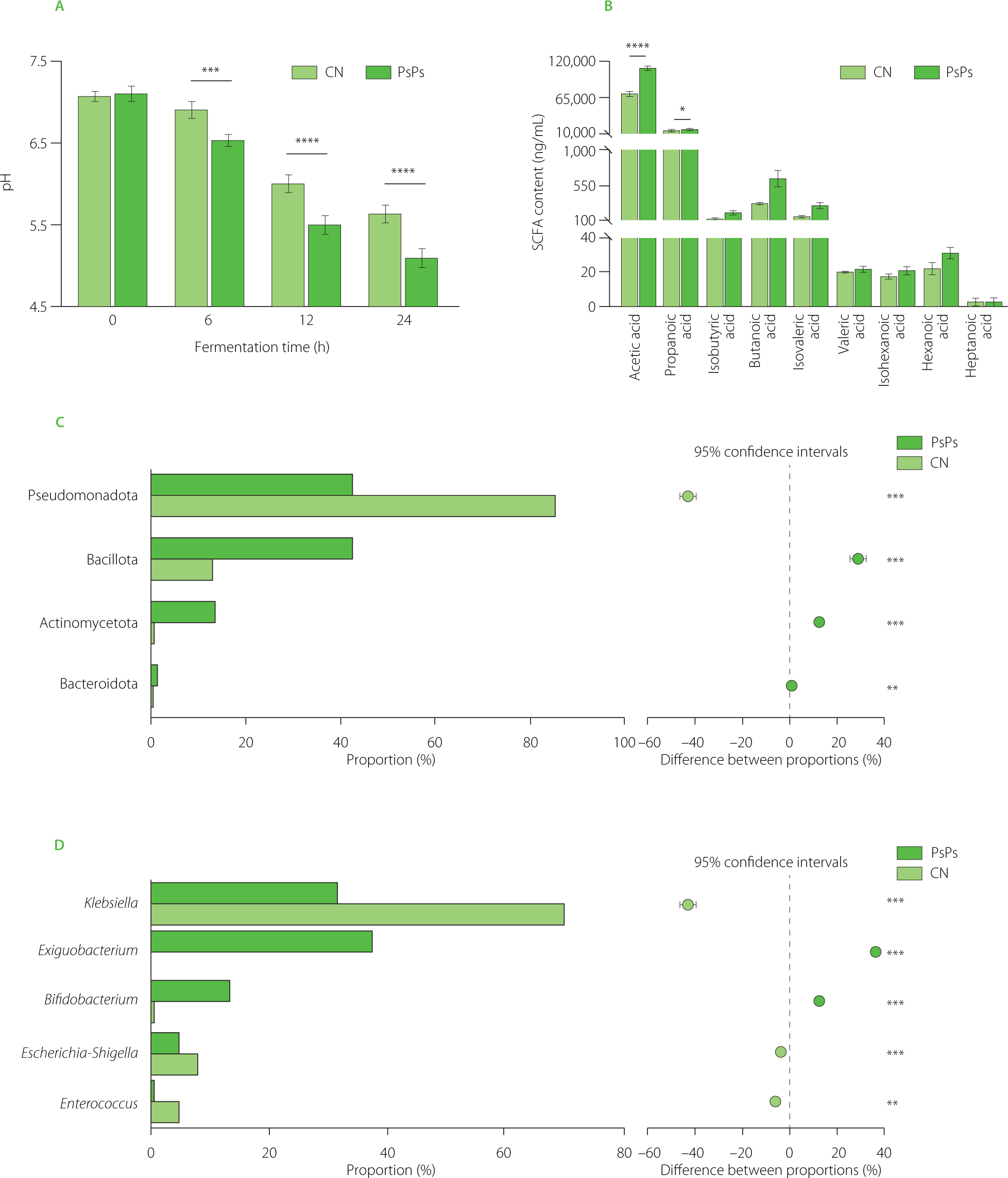
As metabolic byproducts of bacterial fermentation, SCFAs play an essential role in maintaining gut homeostasis and enhancing intestinal barrier function. After 24 h fermentation, the total SCFA level in the PsPs group (131±3 μg/mL) was significantly (p<0.05) higher than in the CN group (86.4±2.1 μg/mL). In addition, the content of both main compounds in the SCFA profile, i.e., acetic acid and propionic acid, was substantially higher in the PsPs than in the CN group (Figure 1B). The elevated levels of acetic acid and propionic acid in the PsPs group were in line with our previous research findings in healthy mice [Luo et al., 2022].
Microbial dynamics in the gut play pivotal roles in regulating host immunity and metabolic health. Figure S2 in Supplementary Materials illustrate changes in the α-diversity of microbial communities in mixed fecal fermentation broth following 24 h fermentation, compared to broth supplemented with PsPs. After PsPs addition, the ACE showed no significant (p≥0.05) change, while the Shannon index increased, and the Simpson index decreased significantly (p<0.05). This indicates that sufficient sample data has been obtained for subsequent analysis. The sample-level hierarchical clustering tree constructed based on ASV levels (Figure S3A in Supplementary Materials) reveals significant intragroup similarities and intergroup differences in microbial community composition between the PsPs and CN groups. The microbial community composition of PsPs and CN at the phylum and genus levels are represented in Figures S3B and C in Supplementary Materials. Comparative analysis of fermentation results between the two sample groups at the phylum level (Figure 1C) demonstrates that PsPs significantly suppressed the Pseudomonadota proportion while significantly increasing the abundance of Bacillota, implying that PsPs have the potency to regulate gut microbiota dysbiosis [Voermans et al., 2025]. PsPs also significantly enhanced the relative abundance of Bacteroidota, and the increase in both Bacteroidota and SCFAs suggests that PsPs may be beneficial for treating dysbiosis [Yu et al., 2023]. At the genus level (Figure 1D), PsPs reduced the abundance of Klebsiella and Escherichia-Shigella, implying its potential to relieve AAD [Li et al., 2023]. It is worth noting that PsPs have a significant promoting effect on Bifidobacterium, which may make them particularly effective in maintaining gut health and mitigating diarrhea symptoms [Hempel et al., 2012]. Overall, PsPs possess prebiotic properties and may provide relief for AAD.
Interestingly, previous studies have shown that PsPs may contain β-glycosidic bonds and branched structures, which protect against degradation by host digestive enzymes while still allowing microbial carbohydrate-active enzymes (CAZymes) to break down [Guan et al., 2026; Subhash et al., 2026]. The interactions among polysaccharide structure, gut microbiota metabolism, and host health are increasingly recognized as the fundamental mechanisms underlying the regulatory effects of polysaccharide prebiotics. Moreover, differences in monosaccharide composition and glycosidic bond configuration determine the selective fermentation patterns and subsequent metabolic outcomes [Zhang et al., 2026]. The selective promotion of specific bacterial groups by PsPs may be related to the action of CAZymes, which are the primary enzymatic systems responsible for polysaccharide degradation in the gut microbiota. Glycosidase hydrolases (GHs) and polysaccharide lyases are organized into polysaccharide utilization loci, enabling gut bacteria to recognize, bind, uptake, and degrade complex polysaccharides [Qu et al., 2025]. The Bifidobacterium enrichment observed during PsPs fermentation is consistent with the characteristic of this genus, possessing multiple CAZymes specialized for the degradation of plant-derived polysaccharides [Pokusaeva et al., 2011]. In detail, the Bifidobacterium possesses numerous glycoside hydrolase (GH) families targeting arabinoxylans and xylans, enabling it to efficiently ferment arabinoxylans and xylans commonly found in plants. Therefore, the significant proliferation effect of Bifidobacterium in PsPs likely reflects the metabolic specialization of Bifidobacterium in utilizing the structure of these polysaccharides, which are rich in arabinose and xylose. The monosaccharide composition of PsPs may partially explain these fermentation changes. Previous research indicates that the monosaccharide composition of dietary fiber influences gut microbiota composition and fermentation products, and that arabinose oligosaccharide structures selectively stimulate beneficial bacteria such as Bifidobacterium [Jensen et al., 2024; Leschonski et al., 2024]. Therefore, the increase in Bifidobacterium and SCFAs may be related to the high levels of xylose and arabinose in PsPs. Furthermore, the presence of minute amounts of galacturonic acid and rhamnose residues may imply the presence of pectin domains, as fermentation of these domains by intestinal pectindegrading bacteria would yield acetate and propionate [Yüksel et al., 2025].
Effects of leaf polysaccharides on mice
After successfully inducing the AAD model in mice, the mice were gavaged with PsPs or a normal saline solution for 7 days. During the gavage period, weights of mice in the AAD and NR groups decreased significantly, whereas in the PsPs group, weights gradually recovered nearing those of the CN group (Figure S4A in Supplementary Materials). Mice in the CN group showed positive behavioral responses, while the mice from the AAD and NR groups experienced evident diarrhea; in contrast, diarrhea symptoms were distinctly alleviated in the PsPs group. This demonstrates that PsPs has an ameliorative effect on AAD in mice. The colon lengths of the four groups of mice were compared (Figure S4B in Supplementary Materials). In AAD and NR groups, the colons were statistically shorter than in both the CN and PsPs groups, while no significant difference was detected between the CN and PsPs groups. This confirms successful modeling of AAD. These recovery patterns are similar to findings from natural polysaccharide studies, which suggest that they may alleviate AAD by restoring microbial fermentation, improving epithelial repair, and limiting inflammatory damage [Lai et al., 2025; Xu et al., 2023]. From a mechanistic perspective, the improvement in body weight, diarrhea symptoms, and colon length demonstrates that PsPs likely accelerate symptom recovery and may also promote the restoration of mucosal homeostasis following antibiotic-induced ecological disruption.
The arrow in the HE–stained images of cecal tissues of the AAD mice (Figure 2A) shows substantial inflammatory cell infiltration, verifying successful model development. In addition, the NR group exhibited more mucus accumulation compared to the PsPs and CN groups, implying that PsPs may possess certain restorative activity against intestinal pathology. The histological differences between the PsPs group and the NR group have potential biological significance, as natural recovery following antibiotic suspension may be insufficient to rapidly rehabilitate mucosal metabolism. Given that intestinal barrier dysfunction and LPS derived from the microbiota can exacerbate systemic inflammation [Di Vincenzo et al., 2024], the attenuated inflammatory infiltration observed in the PsPs group supports a mechanism involving the microbiotabarrier-inflammation axis, not simply a cessation of diarrhea.
Figure 2
(A) Images of hematoxylin and eosin (HE) stained mice cecum (×40 and ×200). (B) Immunofluorescence microscope images of the mice cecum, with brighter fluorescence indicating higher expression levels. AAD, antibiotic-associated diarrhea group; CN, blank control group; PsPs, Polygonatum sibiricum Red. leaf polysaccharides group; NR, natural recovery group.
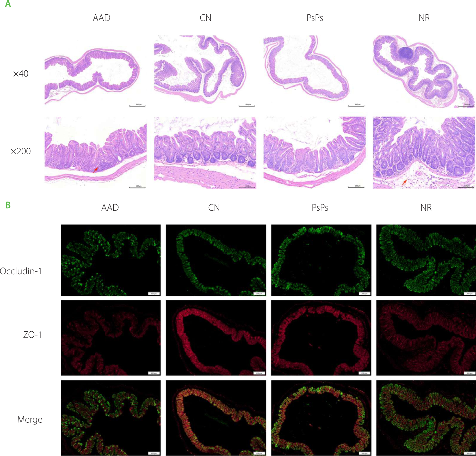
Tight junction proteins are vital for maintaining the integrity of the intestinal barrier. The impact of PsPs on tight junction proteins was investigated through immunofluorescence analysis (Figure 2B). Images with higher brightness indicate relatively stronger expression levels of the relevant indexes. Compared with the AAD and NR groups, PsPs could upregulate the expression of ZO-1 in the intestine, rendering the expression profile closer to the CN group. This demonstrates that PsPs have the capacity to strengthen intestinal barrier functions. It is possible that this is related to the microbial fermentation driven by PsPs.
Effects of leaf polysaccharides on the gut microbiota of mice
The relative abundance and diversity of species in the experimental groups were analyzed with α-diversity statistics. As evidenced in Figure S5 in Supplementary Materials, there were statistical differences in the ACE, Shannon, and Simpson indexes across the four groups, reflecting alterations in the richness and evenness of the gut microbiota in mice following PsPs gavage. PCoA based on ASV levels demonstrated tight clustering of individuals within each group, with significant differences observed in the CN and AAD groups compared to the PsPs or NR groups (Figure 3A), as well as distinct separations between the PsPs and NR groups (Figure 3B). These results suggest that the microbial community composition in the PsPs and NR groups has undergone considerable modification compared to the CN and AAD groups, and furthermore, the microbial structure of the PsPs group differed significantly from the NR group. Moreover, compared with the AAD group, the PsPs group showed significant improvements in microbial richness and evenness.
Figure 3
The effect of PsPs on mice gut microbiota. (A) Plot of principal co-ordinates analysis (PCoA) of gut microbiota in four groups on amplicon sequence variant (ASV) level. (B) Plot of PCoA of gut microbiota in PsPs and NR groups on ASV level. AAD, antibiotic-associated diarrhea group; CN, blank control group; PsPs, Polygonatum sibiricum Red. leaf polysaccharides group; NR, natural recovery group.
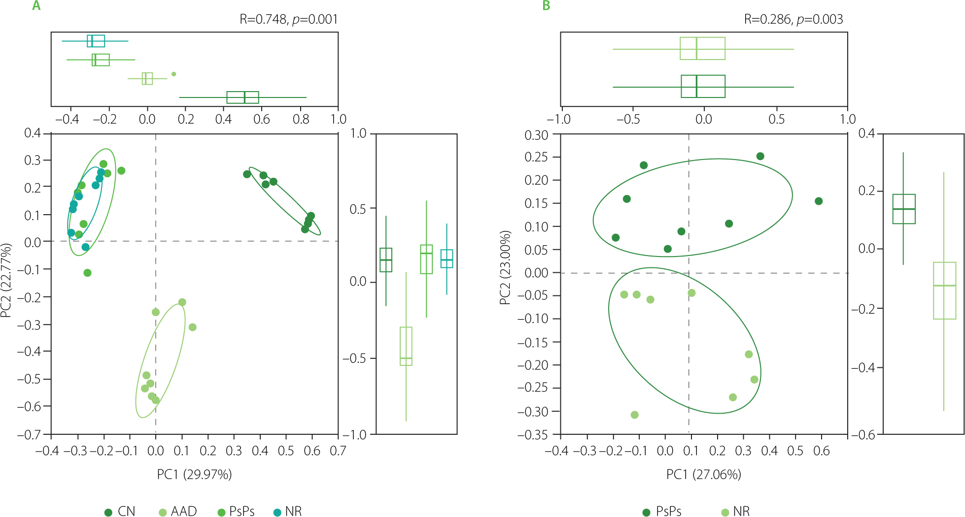
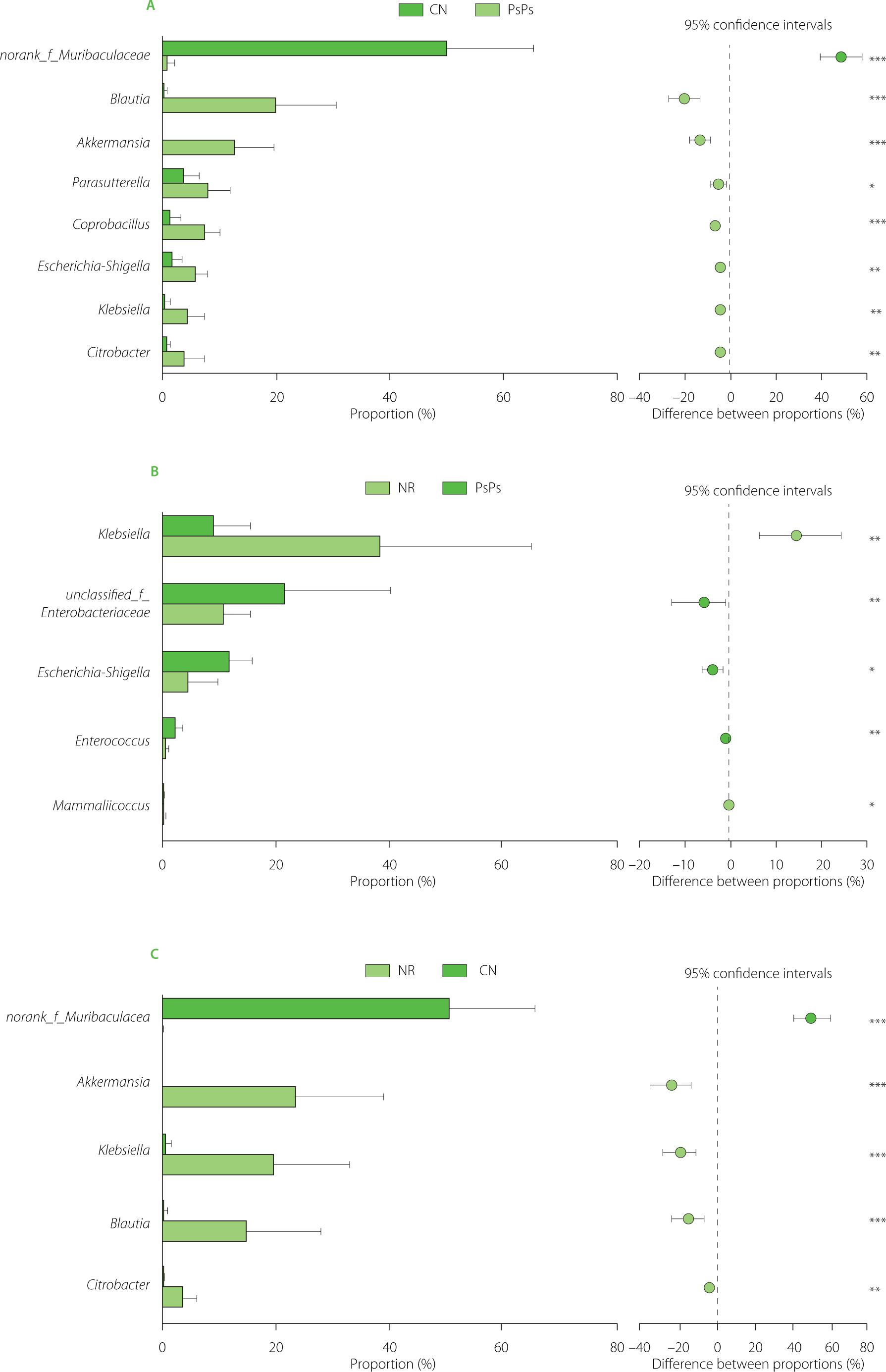
Figure S6 in Supplementary Materials showcase the compositional differences in the gut microbiota across the four groups at the phylum and genus levels. As shown in Figure 4A–C, PsPs notably suppressed Klebsiella abundance compared to the NR; this is likely the primary factor contributing to AAD alleviation. However, in contrast to the outcomes of in vitro fermentation experiments, PsPs did not display an inhibitory effect on the proportion of Escherichia-Shigella in AAD animal experiments. It is particularly noteworthy that the Blautia content was significantly higher in the PsPs group than in the CN group, which may be the major factor facilitating the recovery of cecal tissue in the PsPs group. Blautia is a probiotic that helps balance host health [Liu et al., 2021]. Blautia stimulates immune regulation, and increased concentrations are often accompanied with higher acetate production, which is supported by findings from in vitro fermentation experiments on augmented SCFA yields [Ye et al., 2023]. And compared to the CN group, the richness of Akkermansia was also substantially higher in the PsPs group, which may be another important way PsPs supports gut wellness. These results all demonstrate that PsPs can alleviate AAD and confer prebiotic benefits. In the context of AAD, the selective inhibition of Klebsiella is particularly important, as antibiotic pressure often promotes the proliferation of facultative anaerobes and Enterobacteriaceae. This increases the endotoxin burden and exacerbates epithelial inflammation. Therefore, the diminished Klebsiella levels in the PsPs group suggest that PsPs may have attenuated the inflammation-driving response triggered by pathogen-associated microbiota. The enrichment of Blautia may promote epithelial repair through acetic acid production and mucus maintenance, which aligns with reports that SCFAs sourced from Blautia support colonic mucus function [Holmberg et al., 2024]. The increase in Akkermansia is also noteworthy, as this mucus-associated bacterium is involved in mucus layer renewal as well as host metabolic and immune homeostasis [Ioannou et al., 2025]. Remarkably, because the initial microbial communities in the feces of healthy humans differ from those in mouse feces, this complex ecosystem has prevented the inhibitory effect of PsPs on microbial communities observed through in vitro fermentation, which was not fully reproduced in the disease mouse model. Similar observation has also been made in our previous research [Ma et al., 2025]. Although some microbial changes in the PsPs group tended toward the NR group, they also exhibited distinct differences in overall microbiota composition in the PCoA, suggesting that PsPs administration in mice might not only promote recovery but also increase the proportion of certain beneficial bacteria compared to natural recovery. Taken together, the in vivo microbial profile suggests PsPs may shift the post-antibiotic ecosystem from a state dominated by pathogenic commensals to a microbiota that supports metabolism and barrier function.
Figure 4
Comparison of the mice gut microbiota: (A) CN and PsPs groups, (B) PsPs and NR groups, and (C) CN and NR groups. CN, blank control group; PsPs, Polygonatum sibiricum Red. leaf polysaccharides group; NR, natural recovery group. *, **, and *** indicate statistical significance at p<0.05, p<0.01, and p<0.001, respectively.
Effects of leaf polysaccharides on serum cytokine levels
Inflammatory cytokine levels are closely associated with disease development. Changes in serum cytokine levels in AAD mice after PsPs gavage intervention are shown in Table 3. The concentrations of MCP-1, LPS, IL-6, and TNF-α in the PsPs group were significantly lower than in the AAD and NR group, and there was no obvious difference in the levels of MCP-1, IL-6, and TNF-α between the PsPs and CN group. MCP-1 is intimately connected with numerous inflammatory diseases [Singh et al., 2021]. Raised MCP-1 expression in the AAD group signifies successful model induction, while MCP-1 in PsPs declines to near the CN group level, reflecting an effective attenuation of the inflammatory response triggered by AAD. The elevation of LPS is related to gut microbiota dysbiosis and intestinal barrier dysfunction [Di Vincenzo et al., 2024]. Compared to the AAD and NR group, the LPS contents in the PsPs group were considerably lower, which may be attributed to the fact that PsPs could modulate the microbiota composition and enhance the ZO-1 expression of the intestinal barrier. Overall, the reduction in cytokine levels demonstrates that PsPs possess pronounced anti-inflammatory activity in vivo and can mitigate the inflammatory reaction elicited by AAD. Notably, these responses may be linked to previously observed modifications in gut flora, hinting that PsPs may exercise its immunomodulatory function via the gut-immune axis [Dinakis et al., 2024]. Decreases in MCP-1 levels indicate reduced chemotactic recruitment of monocytes and macrophages, while decreased levels of IL-6 and TNF-α suggest that pro-inflammatory cascades are restrained. Considering the reduced LPS levels and the restoration of ZO-1 staining, these data suggest that PsPs may alleviate systemic inflammation by limiting the transport of endotoxins through the diseased intestinal barrier.
Table 3
Serum cytokine levels in mice from experimental groups.
[i] Data are mean ± standard deviation (n=8). Letter symbols: when the superscript letters in each column are completely different, a significant difference is indicated (p<0.05). MCP-1, monocyte chemoattractant protein-1; LPS, lipopolysaccharide; IL-6, interleukin-6; TNF-α, tumor necrosis factor-alpha; AAD, antibiotic-associated diarrhea group; CN, blank control group; PsPs, Polygonatum sibiricum Red. leaf polysaccharides group; NR, natural recovery group.
These findings also support the potential of PsPs for application in animal nutrition. In poultry, the addition of P. sibiricum polysaccharides to feed enhances growth performance, antioxidant capacity, intestinal structure, and the gut microbiota [Yang et al., 2024]. Therefore, PsPs also hold promise as a lowcost prebiotic feed ingredient; however, their dosage, safety, palatability, and cost-effectiveness for different species require further validation. Nevertheless, this study has certain limitations, such as the use of only 16S rRNA sequencing and the absence of experiments with positive controls. In addition to the perspective of feed composition, future research should transition from static endpoint observations to dynamic intestinal physiological models. “Gut-on-a-chip” platforms can replicate luminal flow, shear stress, mucus secretion, the epithelium-endothelium compartment, immune cell interactions, and peristaltic-like mechanical signals, thereby establishing a controllable bridge between anaerobic fermentation and animal models [Taavitsainen et al., 2024]. A recent platform integrating human fecal microbiota and peristaltic-like movements reveals that multi-omics readouts can be used to replicate and monitor host-microbiome interactions and barrier responses [Ballerini et al., 2025]. For PsPs, such systems will enable real-time assessment of changes in mucus kinetics, cytokine release, and microbial metabolites under antibiotic interference and PsPs intervention. In parallel, complementary multi-omics analyses will provide further insight into the mechanisms underlying the detected 16S rRNA changes. Broad-range metagenomics will determine whether the increase in Blautia and Akkermansia is accompanied by enrichment of CAZymes and SCFAs synthesis pathways, or depletion of the pathogenic bacterium Klebsiella. Targeted and untargeted metabolomics can link microbiome changes to SCFAs and inflammatory lipid mediators, while metabolometranscriptomics or metabolic modeling can reveal whether these pathways are transcriptionally active [Dell’Olio et al., 2024; Go et al., 2024]. This strategy helps distinguish taxa solely associated with recovery from microbial functions that may mediate barrier repair and anti-inflammatory effects.
CONCLUSIONS
This study investigated the alleviating effects of PsPs on AAD mice model and its potential mechanisms. PsPs exhibits gastrointestinal stability in vitro, enables utilization by gut microbiota, promotes the generation of SCFAs through fermentation, and reshapes the microbial community by reducing conditionally pathogenic and disease-causing bacterial genera (such as Klebsiella and Escherichia-Shigella) in vitro, while increasing beneficial or barrier-associated taxa (including Blautia and Akkermansia) in vivo. In mouse models, PsPs alleviated diarrhea-related phenotypes, improved colonic length and cecal histological structure, enhanced ZO-1 expression, and reduced serum levels of MCP-1, LPS, IL-6, and TNF-α. Conclusively, we hypothesize that PsPs may alleviate AAD by modulating the gut microbiota – SCFAs – barrier – inflammation axis, wherein the restoration of microbial fermentation function and the suppression of pathogen-associated endotoxin stress jointly promote epithelial repair and immune rebalancing. This work provides scientific evidence for the highvalue utilization of the non-traditional application part of Polygonatum leaves, and offers new insights toward developing applications of polysaccharides derived from food and medicine homology in the fields of functional foods and pharmaceuticals.