INTRODUCTION
Members of the genus Listeria are Gram-positive, bacteria that are ubiquitous in natural ecosystems as well as in agricultural and food-processing environments. Their ability to tolerate osmotic stress, grow at refrigeration temperatures, and form biofilms enables their persistence in food-processing facilities and along the production chain [Cebeci & Otlu, 2024]. Among the described species, Listeria monocytogenes is the main pathogen responsible for listeriosis in humans and animals [Kaszoni-Rückerl et al., 2020]. In contrast, Listeria innocua has long been considered nonpathogenic and is commonly used as a model organism due to its close genetic relatedness to L. monocytogenes [Matto et al., 2022]. It is frequently isolated from raw materials and food-processing environments, often co-occurring with L. monocytogenes and, in many cases, representing the predominant Listeria species detected [Arslan et al., 2020]. Despite its traditional classification as harmless, recent studies have suggested that certain atypical L. innocua strains may harbor virulence-associated determinants. These strains may arise through horizontal gene transfer or due to evolutionary divergence from a virulent ancestor shared with L. monocytogenes [Ramadan et al., 2023; Zanolli Moreno et al., 2012]. Phylogenetic analyses support this view, indicating that both species originated from a common ancestor, with the attenuated phenotype of L. innocua likely resulting from the loss of key virulence genes [Clayton et al., 2014; Gana et al., 2023]. The presence of pathogenicity islands, such as Listeria pathogenicity island 1 (LIPI-1), (Listeria pathogenicity island 3 (LIPI-3), and Listeria pathogenicity island 4 (LIPI-4), in atypical L. innocua isolates further supports their evolutionary and genetic relatedness to L. monocytogenes [Clayton et al., 2014; Li M. et al., 2021; Zanolli Moreno et al., 2012]. Although these strains generally exhibit lower virulence than L. monocytogenes [Moura et al., 2019], some hemolytic variants have demonstrated the ability to cross the intestinal barrier and disseminate within host tissues [Gwida et al., 2020]. Clinical reports have linked atypical L. innocua to severe infections in immunocompromised individuals, including bacteremia [Perrin et al., 2003], meningitis [Favaro et al., 2014], ventriculoperitoneal shunt infection [Karli et al., 2014], neonatal listeriosis [Arumugam et al., 2021], meningoencephalitis [Liao et al., 2022], and joint infection [Gupta et al., 2024]. Animal infections, such as cerebral listeriosis in cattle, have also been described [Matto et al., 2022; Rocha et al., 2013].
Virulence in pathogenic Listeria species is mediated by a coordinated set of adhesion, invasion, and intracellular survival factors enabling colonization of the gastrointestinal tract and translocation across the intestinal barrier [Wiktorczyk-Kapischke et al., 2023]. The core virulence determinants are primarily located within LIPI-1, which comprises genes encoding two phospholipases (plcA and plcB), the pore-forming toxin (hly), a metalloprotease (mpl), and the actin-polymerization factor (actA) responsible for intracellular motility. Expression of LIPI-1 genes is regulated by the transcriptional activator PrfA (encoded by prfA gene) [Lee et al., 2023]. Among the genes located within LIPI-1, the most critical is hly, which encodes listeriolysin O (LLO), a toxin responsible for β-hemolytic activity. L. innocua, which typically lacks the hly gene, is therefore generally considered a nonhemolytic species. However, atypical L. innocua strains exhibiting hemolytic activity have also been described [Johnson et al., 2004]. Conversely, rare, atypical L. monocytogenes strains, in which spontaneous loss of virulence genes resulted in a nonhemolytic phenotype, have been reported by Olaimat et al. [2018]. These observations highlight the necessity for continuous monitoring of hemolytic properties and associated virulence determinants. Another important group of virulence-associated proteins are internalins, i.e., surface proteins mediating adhesion and invasion of host cells. The best-characterized members include inlA and inlB, while additional internalins, such as inlC, inlJ, inlH, inlK, inlL, inlF, and inlP, have also been implicated in host interaction and dissemination [Liu et al., 2007]. Other factors contributing to pathogenic potential include the invasion-associated protein (iap), flagellin (flaA), and the alternative sigma factor B (sigB), which regulates the general stress response and enhances bacterial survival under adverse environmental conditions [Osek & Wieczorek, 2022]. In addition to LIPI-1, hypervirulent Listeria strains may harbor LIPI-3 and LIPI-4. LIPI-3 consists of eight genes (llsA, llsG, llsH, llsX, llsB, llsD, llsP, and llsY) encoding listeriolysin S (LLS), i.e., a second cytolytic peptide that exhibits properties of both a bacteriocin and a hemolytic cytotoxic factor. LLS contributes to the modulation of the intestinal microbiota and promotes gastrointestinal colonization [Clayton et al., 2014; Cotter et al., 2008; Lee et al., 2023]. Notably, the llsX gene is commonly regarded as a molecular marker for the presence of the LIPI-3 [Wiktorczyk-Kapischke et al., 2023]. LIPI-4, composed of six genes, encodes a cellobiose-family phosphotransferase system (PTS) and has been associated with enhanced neural and placental tropism, suggesting a role in central nervous system and maternal-neonatal infections [Lee et al., 2023; Li M. et al., 2021]. Although virulence factors have been extensively investigated in L. monocytogenes, knowledge regarding their distribution in other Listeria species remains limited.
Listeriosis remains a significant cause of mortality worldwide. Identifying and characterizing virulence factors is essential for both basic scientific research and ongoing medical challenges. Therefore, this study aimed to investigate the occurrence of selected virulence-associated genes and the hemolytic phenotype of L. innocua isolates recovered from meat products and meat-processing environments in Poland.
MATERIALS AND METHODS
Bacterial isolates and genetic material
A total of 51 L. innocua isolates were included in this investigation. Of these, 32 were recovered from raw poultry, pork, and beef, as well as from processed meat products, including bacon, ham, and sausages. The remaining 19 isolates originated from meat-processing environments and were recovered from swab samples collected from production lines, processing equipment, walls, and floors. The isolates were obtained through collaboration with an accredited microbiological diagnostic laboratory in Poland. Due to the confidential nature of the source information, detailed data regarding the exact origin of the isolates and the individual meat-processing facilities cannot be disclosed. All isolates were obtained in Poland during 2020–2021. Genomic DNA was extracted with the Genomic Mini kit (A&A Biotechnology, Gdańsk, Poland) following the supplier’s protocol and served as the template for PCR analyses. Isolates stored at −80°C in brain heart infusion (BHI; Oxoid, Warsaw, Poland) supplemented with glycerol were used for the evaluation of hemolytic activity. Prior to the experiments, the taxonomic identity of all isolates had been verified as L. innocua via polymerase chain reaction–restriction fragment length polymorphism (PCR-RFLP) and multiplex polymerase chain reaction (multiplex PCR). Exact methodologies were reported previously [Li F. et al., 2021; Paillard et al., 2003].
Detection of virulence-associated genes
Conventional PCR was performed to detect selected virulence-associated genes in L. innocua isolates. The analysis of the actA gene was conducted using two primer pairs, designated actA1 and actA2. Additionally, PCR assays targeting LIPI-4 genes were subjected to optimization; reproducible amplification was achieved for licC and glvA, whereas optimization attempts for the remaining genes did not yield reproducible results across all isolates. Therefore, these genes were excluded from further analysis. Each PCR reaction contained 0.2 U of RUN DNA polymerase (A&A Biotechnology) with the corresponding reaction buffer, 0.2 mM of each deoxynucleoside triphosphate (dNTP) (A&A Biotechnology), and the appropriate primer pair (Table 1), synthesized by Genomed S.A. (Warsaw, Poland). Approximately 10 ng of template DNA were added to each reaction, and the final reaction volume was adjusted to 10 μL. PCR amplification was carried out using a Biometra TOne thermal cycler (Biometra, Göttingen, Germany). Three L. monocytogenes isolates obtained from food samples were used as positive controls. Following amplification, PCR fragments were examined by agarose gel electrophoresis using Midori Green Advance (1×; NIPPON Genetics EUROPE, Düren, Germany). Gel images were acquired with the Gel Doc imaging system (Bio-Rad, Hercules, CA, USA). For each target gene, a representative fragment of the expected length was selected for purification with the Gel-Out Concentrator kit (A&A Biotechnology) and subsequently subjected to Sanger sequencing by Genomed S.A. (Warsaw, Poland). The resulting sequences were compared against reference entries in the NCBI BLAST database (version 2.15.0) to verify the identity of the amplified targets.
Table 1
Detailed information about PCR conditions.
| Virulence genes | Primers name | Target | Primers’ sequence | Annealing temperature (°C) | Amplicon length (bp) | Reference |
|---|---|---|---|---|---|---|
| Listeria pathogenicity island 1 (LIPI-1) | prfA | Listeriolysin-positive regulatory protein | F: 5′-GATACAGAAACATCGGTTGGC-3′ R: 5′-GTGTAATCTTGATGCCATCAGG-3′ | 49 | 274 | D’Agostino et al. [2004] |
| plcA | Phosphatidylinositol-specific phospholipase C | F: 5′-CTGCTTGAGCGTTCATGTCTCATCCCCC-3′ R: 5′-CATGGGTTTCACTCTCCTTCTAC-3′ | 60 | 1,484 | Notermans et al. [1991] | |
| plcB | Phosphatidylicholin-specific phospholipase C | F: 5′-GCAAGTGTTCTAGTCTTTCCGG-3′ R: 5′-ACCTGCCAAAGTTTGCTGTGA-3′ | 55 | 795 | Nishibori et al. [1995] | |
| mpl | Metalloprotease | F: 5′-GGCTCATTTCACTATGACGG-3′ R: 5′-GCTTCCCAAGCTTCAGCAACT-3′ | 55 | 1,473 | ||
| hly | Listeriolysin O | F: 5′-GCAGTTGCAAGCGCTTGGAGTGAA-3′ R: 5′-GCAACGTATCCTCCAGAGTGATCG-3′ | 60 | 210 | Park et al. [2006] | |
| actA1 | Actin polymerization protein | F: 5′-CGCCGCGGAAATTAAAAAAAGA-3′ R: 5′-ACGAAGGAACCGGGCTGCTAG-3′ | 62 | 839 (or 950) | Jallewar et al. [2007] | |
| actA2 | Actin polymerization protein | F: 5′-GACGAAAATCCCGAAGTGAA-3′ R: 5′-CTAGCGAAGGTGCTGTTTCC-3′ | 63 | 268 (or 385) | Jaradat et al. [2002] | |
| Other virulence-associated genes | sigB | Sigma factor | F: 5′-TCATCGGTGTCACGGAAGAA-3′ R: 5′-TGACGTTGGATTCTAGACAC-3′ | 51 | 310 | Bae et al. [2012] |
| iap | Invasion associated protein | F: 5′-ACAAGCTGCACCTGTTGCAG-3′ R: 5′-TGACAGCGTGTGTAGTAGCA-3′ | 56 | 131 | Furrer et al. [1991] | |
| flaA | Flagellin | F: 5′-TTACTAGATCAAACTGCTCC-3′ R: 5′-AAGAAAAGCCCCTCGTCC-3′ | 54 | 538 | Borucki & Call [2003] | |
| Internalins | inlA | Internalin A | F: 5′-ACGAGTAACGGGACAAATGC-3′ R: 5′-CCCGACAGTGGTGCTAGATT-3′ | 55 | 800 | Liu et al. [2007] |
| inlJ | Internalin J | F: 5′-TGTAACCCCGCTTACACAGTT-3′ R: 5′-AGCGGCTTGGCAGTCTAATA-3′ | 55 | 238 | ||
| inlB | Internalin B | F: 5′-CATGGGAGAGTAACCCAACC-3′ R: 5′-GCGGTAACCCCTTTGTCATA-3′ | 57 | 500 | Zhang & Knabel [2005] | |
| inlC | Internalin C | F: 5′-CCCACAATCAAATAAGTGACCTT-3′ R: 5′-CTGGGTCTTTGACAGTATTTGTT-3′ | 57 | 400 | ||
| Listeria pathogenicity island 3 (LIPI-3) | llsX | Listeriolysin L | F: 5′-TTATTGCATCAATTGTTC TAGGG -3′ R: 5′-CCCCTATAAACATCATGCTAGTG-3′ | 55.5 | 200 | Vilchis-Rangel et al. [2019] |
| llsA | F: 5′-CGATTTCACAATGTGATAGGATG-3′ R: 5′-GCACATGCACCTCATAAC-3′ | 52 | 280 | Clayton et al. [2014] | ||
| llsG | F: 5′-GAGACTGGGCTTACTTGC-3′ R: 5′-TACCTCCTGTTCACTGCTTG-3′ | 50 | 415 | Zhang et al. [2019] | ||
| llsH | F: 5′-ATGATGTTCGCTATGGTT-3′ R: 5′-ACATTCCTACTGGCATCA-3′ | 45 | 421 | |||
| llsB | F: 5′-TTACAATCAACCACCAGG-3′ R: 5′-AGTGAACCGAATGACAGA-3′ | 45 | 334 | |||
| llsD | F: 5′-TATGGTGGTATGGAGGGT-3′ R: 5′-ATCACCCTGCTTATTTCA-3′ | 45 | 562 | |||
| llsP | F: 5′-TTTCCAGGTATGCTTCTT-3′ R: 5′-CAATTACGGTGGTTCTCA-3′ | 45 | 554 | |||
| llsY | F: 5′-ATTAGAATAGGAACGCAGAC-3′ R: 5′-TCATAGCACCCAGTTTCG-3′ | 50 | 581 | |||
| Listeria pathogenicity island 4 (LIPI-4) | licA | Phosphotransferase system (PTS) sugar transporter enzyme IIA (EIIA) subunit | F: 5′-GCCTCTTCCTCGTTTCTA-3′ R: 5′-GACTTAACTAAATCGCAGTA-3′ | 45 | 227 | |
| licB | Phosphotransferase system (PTS) sugar transporter enzyme IIB (EIIB) subunit | F: 5′-ATTGCGGCATCTGAGAAA-3′ R: 5′-CAGCGATTAGAATTGGTACTGC-3′ | 55 | 232 | ||
| licC | Phosphotransferase system (PTS) sugar transporter enzyme IIC (EIIC) subunit | F: 5′-GGGATTCCGAAACTACCT-3′ R: 5′-CGAGTGCTCCTGTAACCC-3′ | 47 | 736 | ||
| Lm900558- 70012 | Phosphotransferase system (PTS)-associated protein | F: 5′-TGGTAACAATGCCTGCTT-3′ R: 5′-GCTGAAAGCCCACTGTAT-3′ | 50 | 369 | ||
| Lm900558- 70013 | Transcriptional antiterminator | F: 5′-TATTCAGTGGTTACGAGGCT-3′ R: 5′-CTCCGCCGAAATCTGGTA-3′ | 50 | 403 | ||
| glvA | Maltose-6′-phosphate-glucosidase | F: 5′-TTACTATTGCTGGCGGAGGA-3′ R: 5′-TGCTCACGACCATCCATT-3′ | 47 | 847 |
Hemolysis assay
Hemolytic activity was assessed using Columbia Agar supplemented with 5% sheep blood (Oxoid, Warsaw, Poland). Bacterial isolates stored as glycerol stocks were first streaked onto BHI agar and incubated at 35°C for 24 h. Subsequently, cultures were re-streaked onto blood agar plates and incubated at 35°C for 24 h and 48 h, with hemolysis evaluated after each incubation period. Listeria ivanovii ATCC 19119 was used as a positive control.
RESULTS AND DISCUSSION
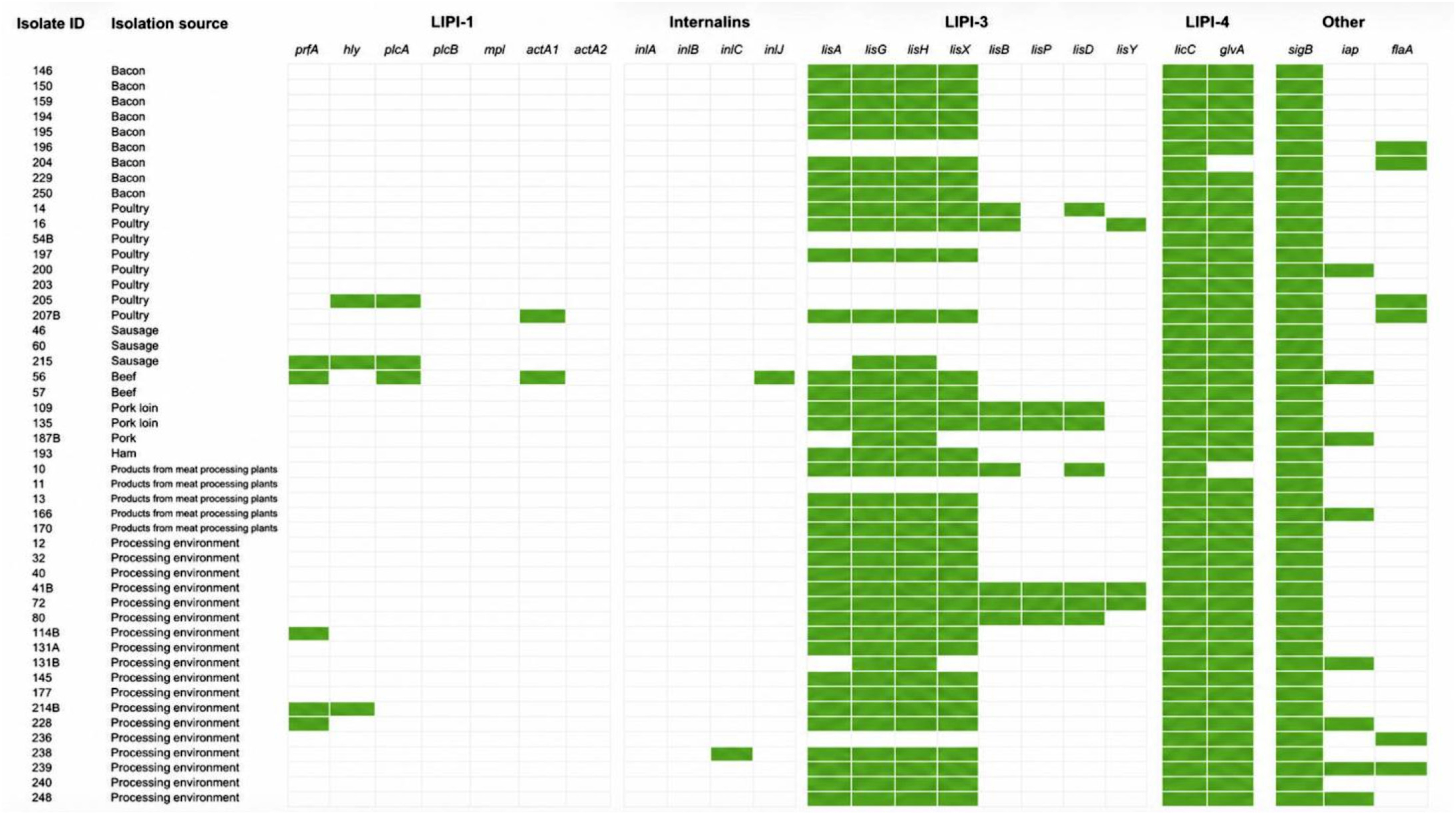
Listeriosis is a significant foodborne disease transmitted primarily through contaminated food, including meat and meat products [Zanolli Moreno et al., 2014]. While the pathogenicity of L. monocytogenes is well characterized in terms of its virulence gene [Freitag et al., 2009], the virulence potential of non-pathogenic Listeria species remains poorly understood. Therefore, this study analyzed the presence of 28 virulence-associated genes in 51 L. innocua isolates by PCR. Detailed results regarding the presence of individual virulence-associated genes in each isolate, together with the corresponding source of isolation, are presented in Figure 1.
Figure 1
Presence of selected virulence-associated genes among Listeria innocua isolates from meat, meat products, and meat-processing environments analyzed in this study. Colored boxes indicate the presence (green) or absence (white) of each gene.
Presence of LIPI-1 genes and internalin-associated genes
Genes associated with the LIPI-1 were detected sporadically among the analyzed isolates (Figure 1). The prfA gene was identified in 5 isolates (10%) from raw meat samples and food-processing environments, while plcA and hly were each detected in 3 isolates (6%). The actA1 gene was present in 2 isolates (4%), whereas plcB, actA2, and mpl were not detected in any strain. In contrast, the stress-response regulator sigB, which is not part of LIPI-1, was present in all isolates (100%). Additionally, the iap gene was identified in 7 isolates (14%), and flaA in 6 isolates (12%). Notably, none of the analyzed isolates carried a complete LIPI-1.
The prevalence of LIPI-1 genes observed in the present study was generally lower than that reported in several previous investigations. For example, plcA was detected in 97.4% of L. innocua isolates recovered from beef, fish, raw milk, and traditional cheese samples in Turkey, followed by iap (35.8%) and hlyA (15.3%) [Cebeci et al., 2024]. In the present study, plcA and hly were identified in only 6% of the isolates, while iap was detected in 15%, indicating a substantially lower prevalence of these virulence-associated determinants. The occurrence of the iap gene appears to vary considerably among studies. A markedly higher prevalence was reported, with iap identified in 36 isolates from the poultry production chain [Gwida et al., 2020]. Similarly, the gene was detected in isolates originating from cattle and sheep [Matto et al., 2022], raw milk and yogurt samples [Ramadan et al., 2023] and grain samples [Yong et al., 2024]. The prevalence of plcA and hly has been reported to vary considerably among L. innocua isolates, with one study detecting both genes in all five isolates recovered from swine slaughterhouses and meat markets, whereas another identified them in only 9 of the 42 isolates collected from fecal samples of healthy birds in Finland [Moura et al., 2019; Zanolli Moreno et al., 2014]. In Polish research, hly was identified in 16.7% of isolates from fish and shrimp, while plcA and iap were absent [Zakrzewski et al., 2020]. In contrast, no presence of hly, plcA, or iap was detected in L. innocua isolates from food and food-contact surfaces [Mafuna et al., 2022; Rossi et al., 2020]. The discrepancies between studies may reflect differences in isolate origin, genomic diversity, and the dynamic nature of virulence-associated gene acquisition or loss within L. innocua populations.
The present study demonstrated a very limited distribution of internalin-associated genes among the analyzed L. innocua isolates (Figure 1). Neither inlA nor inlB was detected in any of the examined strains. In contrast, inlC was identified in a single isolate recovered from a food-processing environment, while inlJ was detected in one isolate originating from a beef sample (2% each).
Similarly, low prevalence of internalin genes has been reported in other studies. In the study, five atypical L. innocua isolates were described, all of which harbored inlA and inlJ, suggesting that certain atypical strains may carry a broader repertoire of virulence-associated genes [Zanolli Moreno et al., 2012]. The presence of inlA in only a single isolate obtained from food and food-manufacturing plants has also been reported [Rossi et al., 2020]. Conversely, no inlA, inlB, inlC, inlJ, or inlK genes were detected among isolates from the poultry production chain [Gwida et al., 2020]. Other studies have also reported the absence of major virulence determinants in L. innocua. Isolates from fresh and frozen soft fruits lacked LIPI-1 and internalin genes [Wartha et al., 2023], while strains from slaughterhouses were found to be devoid of LIPI-1, internalins, and LIPI-3 elements [Qi et al., 2024].
Although LIPI-1 and internalin-associated genes were generally detected sporadically and most often occurred individually, two isolates stood out due to their notably expanded virulence gene profiles. Isolate ID56, recovered from beef, harbored six virulence-associated genes (prfA, sigB, iap, plcA, actA, and inlJ), representing one of the most complex profiles identified in this study. Similarly, isolate ID 205, originating from poultry, carried four virulence-associated genes (sigB, plcA, hly, and flaA). The presence of multiple virulence determinants within single isolates is noteworthy and may indicate an increased adaptive or pathogenic potential, warranting further investigation to assess their functionality and possible contribution to virulence.
Presence of LIPI-3 genes
In contrast to the limited distribution of LIPI-1 and internalin-associated genes, LIPI-3 genes were considerably more prevalent among the analyzed L. innocua isolates. Genes located in the 5′ region of the LIPI-3 were detected at high frequencies. The llsA and llsX genes were each identified in 36 isolates (69%) and consistently co-occurred within the same strains. Similarly, llsG and llsH were detected in 37 isolates (73%) and were always present together. Notably, all isolates harboring llsA and llsX also carried llsG and llsH, indicating a strong tendency for cooccurrence of genes located in the 5′ region of the island. In contrast, genes located in the 3′ region of LIPI-3 were detected much less frequently. The llsB and llsD genes were each identified in 7 isolates (13%) and consistently co-occurred. The llsP gene was detected in 5 isolates (10%), whereas llsY was found in only 3 isolates (6%). A complete LIPI-3, defined as the presence of all eight analyzed LIPI-3 genes, was identified in 2 isolates (4%), both originating from distinct food-processing environment samples. Partial LIPI-3 profiles, characterized by the presence of at least one LIPI-3 gene, were detected in 40 isolates (78%), whereas 11 isolates (21%) did not harbor any of the analyzed LIPI-3 genes.
The predominance of LIPI-3 genes observed in the present study is consistent with previous findings demonstrating that these genes are more prevalent in L. innocua than other pathogenicity islands. Clayton et al. [2014] analyzed a total of 64 L. innocua isolates recovered from food products, of which 45 strains (70.3%) were positive for llsA. Eleven isolates carried a complete LIPI-3, whereas 25 exhibited a truncated LIPI-3 profile lacking llsB, llsY, llsD, and llsP, corresponding to the presence of llsA, llsG, llsH, and llsX. Nine isolates harbored only llsA, and 19 strains lacked all LIPI-3 genes. Notably, the LIPI-3-positive isolates described in that study were non-hemolytic, consistent with the findings of the present study [Clayton et al., 2014]. In another investigation focusing on non-pathogenic Listeria spp. isolated from food and food-processing facilities, three L. innocua isolates harboring a complete LIPI-3 were identified among 36 analyzed strains [Mafuna et al., 2022]. A larger survey conducted in South Africa examined 110 L. innocua isolates originating from cattle farms, beef abattoirs, and retail outlets. The llsA (66 isolates), llsG (66 isolates), llsH (65 isolates), and llsX (65 isolates) genes were frequently detected, whereas genes located in the 3′ region- llsB (7 isolates), llsD (8 isolates), llsP (6 isolates), and llsY (8 isolates), were considerably less prevalent [Gana et al., 2023]. These findings corroborate the results of the present study, confirming the broader distribution of LIPI-3 compared with other pathogenicity islands in L. innocua. Hence, a consistent pattern emerges across studies, including the present one, demonstrating higher prevalence and frequent co-occurrence of genes located in the 5′ region of the island, accompanied by reduced detection of genes positioned toward the 3′ end. The structure of LIPI-3, therefore, appears to vary among L. innocua isolates, ranging from complete islands to partial remnants or total absence, reflecting ongoing genomic variability within the species. Notably, LIPI-3 has been reported to be considerably more widespread in L. innocua than in L. monocytogenes, where it is generally confined to selected hypervirulent lineages [Lee et al., 2023; Zhang et al., 2019].
Presence of selected LIPI-4 genes
Among the LIPI-4 genes, licC and glvA were successfully analyzed. The licC gene was detected in all examined isolates (51/51; 100%), while glvA was identified in 48 isolates (94%). The remaining LIPI-4 genes were not included in the present analysis.
LIPI-4 is a relatively recently described pathogenicity island and remains the least extensively investigated among the known Listeria pathogenicity islands. In L. monocytogenes, its occurrence is largely restricted to the hypervirulent clonal complex CC4 [Lee et al., 2023]. Data regarding its distribution in L. innocua remains limited compared with LIPI-1 and LIPI-3. Structurally, LIPI-4 encodes a PTS system: licC, encoding the enzyme IIC component (EIIC) of the phosphotransferase system (PTS) sugar transporter, responsible for substrate recognition and facilitates sugar transport into the cell, and glvA, encoding maltose-6′-phosphate glucosidase [Zhang et al., 2019]. Both genes were highly prevalent among the examined isolates, with licC detected in all strains and glvA in the vast majority. Although genes associated with the LIPI-4 region were frequently detected, the limited number of target genes successfully amplified did not allow confirmation of the presence of a complete and functional LIPI-4 in the studied collection. Previous studies have reported the occurrence of intact LIPI-4 in L. innocua, including three food-derived isolates harboring the complete cluster [Li M. et al., 2021], as well as a high prevalence of the full island among 36 analyzed strains [Mafuna et al., 2022]. The frequent detection of licC and glvA suggests enhanced metabolic adaptability of the analyzed isolates, particularly in the utilization of specific carbohydrates. Such capabilities may support persistence in food environments, improve competitiveness against other microorganisms, and potentially facilitate host colonization. At the same time, the widespread presence of these genes cautiously indicates that LIPI-4 related elements may be common in L. innocua. However, further investigations are necessary to determine the presence of the remaining LIPI-4 genes and to clarify the functional significance of the detected determinants.
Hemolysis assay
No hemolytic activity was observed among the analyzed L. innocua isolates, despite the presence of the hly gene in three isolates (205, 214B, and 215), indicating a lack of functional expression of this virulence determinant under the tested conditions. The discordance between the presence of hly and the absence of a clear hemolytic phenotype represents a particularly intriguing finding. Several explanations may account for this observation, including mutations within the hly coding sequence impairing listeriolysin O functionality, nonsense mutations leading to truncated protein products, or alterations within the promoter region affecting gene transcription. Additionally, impaired activity or mutations in the transcriptional regulator prfA could disrupt proper activation of LIPI-1 genes. To clarify the molecular basis of this discrepancy, further investigations should include sequencing of the hly gene and its regulatory regions, as well as analyses of gene expression. Although L. innocua is generally regarded as a non-hemolytic species, hemolytic isolates have occasionally been reported. An atypical strain carrying a complete set of LIPI-1 genes exhibited hemolytic activity but failed to induce disease in murine models, suggesting that hemolysis alone does not necessarily confer full pathogenic potential [Johnson et al., 2004]. In contrast, other studies demonstrated that hemolytic L. innocua isolates harboring LIPI-1 and inlA may exhibit virulence, including the ability to cross the intestinal epithelium and disseminate systemically, albeit to a lesser extent than L. monocytogenes EGDe [Moura et al., 2019]. Weakly hemolytic hly-positive isolates and additional hemolytic LIPI-1 positive strains from pork, seafood, and poultry have also been described [Milillo et al., 2012; Volokhov et al., 2007; Zanolli Moreno et al., 2014]. Although exposure to atypical hemolytic L. innocua strains appears to be rare, both the present findings and previous reports indicate that phenotypic assessment of hemolysis may not always provide reliable results. The emergence of atypical isolates, including strains exhibiting low-hemolytic activity, further complicates species identification when conventional methods are applied. The present findings highlight that the presence of the hly gene alone is not sufficient to infer hemolytic phenotype in L. innocua, emphasizing the importance of combining genotypic and phenotypic approaches in virulence assessment.
CONCLUSIONS
This study provides the first comprehensive characterization of 28 virulence-associated genes in L. innocua isolates recovered from raw and processed meat products as well as meat-processing environments in Poland. The findings indicate a sporadic occurrence of genes clustered within the LIPI-1 and internalin-associated genes. In contrast, LIPI-3 genes, present either as a complete island or in truncated forms, were more widespread, demonstrating a predominance compared with other pathogenicity islands. The universal presence of sigB, together with the frequent detection of licC and glvA, may suggest enhanced metabolic adaptability of the analyzed isolates and could support their persistence in food and food-processing environments. Although L. innocua is generally regarded as non-pathogenic, its occurrence in food matrices warrants attention, particularly due to its frequent coexistence with L. monocytogenes and its potential role as a reservoir of virulence-associated genes. Importantly, the detection of virulence-associated genes at the genomic level does not necessarily imply their functional expression or pathogenic potential. As the present study was limited to DNA-based identification of these determinants, further investigations focusing on transcriptional activity and functional analyses are required to more accurately assess their potential contribution to the pathogenicity of L. innocua. Furthermore, the implementation of whole-genome sequencing (WGS) in future studies could further strengthen this approach by providing comprehensive information on the genetic background, virulence-associated genes, and evolutionary relationships among L. innocua isolates. These findings emphasize the importance of including L. innocua in food safety monitoring and risk assessment frameworks.